Nuclear Medicine
Why Us?
Choithram Hospital & Research Centre has been a pioneer in the field of Nuclear Medicine in Madhya Pradesh since 1983. Our Nuclear Medicine Department stands out for its ability to provide functional information about organs to clinicians, enabling early diagnosis and differentiation between viable, ischemic, and non-viable tissue. With state-of-the-art technology and expertise, we offer safe, non-invasive, and cost-effective screening procedures, ensuring optimal patient care and outcomes.
Highlights of the Department:
Pioneering Legacy: With a history dating back to 1983, Choithram Hospital & Research Centre has been at the forefront of Nuclear Medicine in Madhya Pradesh, providing advanced diagnostic and therapeutic services to patients.
Functional Information: Nuclear Medicine procedures offer functional information about organs, allowing for early detection of diseases and differentiation between different tissue types, providing valuable insights for clinicians.
Safe and Non-Invasive: Our procedures are non-invasive, safe, and involve minimal radiation exposure, making them suitable for patients of all ages. Radiopharmaceuticals used are physiologically inert and are eliminated from the body without retention.
Latest Technology:
Our Nuclear Medicine Department is equipped with state-of-the-art technology, including:
Discovery NM630 SPECT Dual Head Gamma Camera Computer System: This high-tech system offers advanced scanning technology designed to optimize image clarity and sharpness while minimizing scan times and radiation doses. It provides superb image quality and efficient workflow, ensuring accurate diagnoses and improved patient experience.
Discovery IQe 16 Slice PET-CT scanner system- The state-of-the-art imaging technology that matches both metabolic & structural information for accurate diagnosis & treatment planning of cancer patients.
Our Services:
Our department provides a comprehensive range of Nuclear Medicine services, including:
Diagnostic Imaging Modalities:
– Positron Emission Tomography (PET) and Computed Tomography (CT) Scan
– Thyroid Scan
– Iodine 131 Whole-Body Imaging
– Myocardial Perfusion Imaging
-Gated Blood Pool Radionuclide Ventriculography (MUGA)
-Lung Perfusion Scan
-Liver Spleen Scan
– Gastric Emptying Time Evaluation
– Hepatobiliary Scan
– Renal Scan- DTPA, DMSCA, EC Scans
– Bone Scan
Therapeutic Treatment Modalities
– Low Dose Radioactive Iodine Therapy for Thyrotoxicosis
Meet Our Doctors:
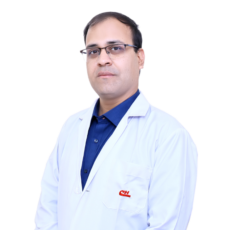
Meet Our Doctors


